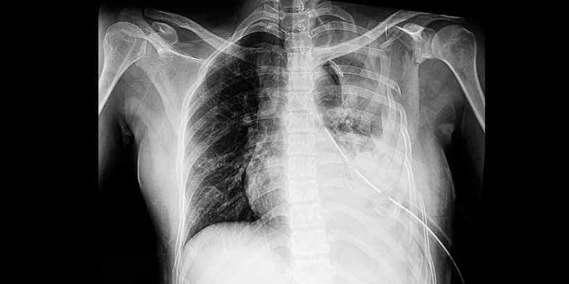
Pulmonary Embolus
 Pulmonary embolism (PE) can be defined as a substance—which arises elsewhere in the body and travels via the bloodstream—blocking the flow of blood within the pulmonary arterial vasculature. (Technically, this makes the term “thromboembolism” more accurate, but clinically the terms are freely interchanged.1) Acute PE is a major cause of morbidity and mortality, with an incidence of 60-70 cases per 100,000 population and associated 30-day all-cause mortality rates between 9% and 11%.6 Recurrent PE events are common, with a cumulative prevalence of up to 30% at 10 years after PE diagnosis; over the long term, chronic thromboembolic pulmonary hypertension (CTEPH) may occur, with an annual incidence of up to 9% at two years after PE diagnosis.6
PE may present with classical features such as breathlessness and pleuritic chest pain, but also less characteristically, such as insidious onset breathlessness over days-to-weeks or syncope with relatively few respiratory symptoms.8 Dyspnea may be followed by chest pain, cough, or hemoptysis.11 Concurrent leg pain may be indicative of concomitant deep vein thrombosis.11 Less common presentations of PE can include arrhythmias, syncope, significant hypoxemia, and hemodynamic collapse.2 The populations at greatest risk of PE include those who had recent trauma, had recent major surgery, are obese, are pregnant, underwent hormonal therapy, smoke, and have certain cancers.7 Clinicians need to have a high degree of suspicion for PE in patients presenting with potential cardiopulmonary symptoms, since the consequences of missing or delaying the diagnosis of PE can be severe.4
The diagnosis of a PE is achieved through clinical evaluation, laboratory testing, electrocardiogram (EKG), radiologic examinations, and echocardiography.1
A common tool used for clinical evaluation of the patient is the Wells criteria scoring system. While there are several clinical probability scores, the Wells score remains the predominant score in international guideline algorithms.4
| Predictor | Score |
| Clinical signs/symptoms of DVT | +3 |
| Alternative diagnosis is less likely | +3 |
| Heart rate > 100 | +1.5 |
| Immobile x 3 days or surgery in last 4 weeks | +1.5 |
| Previous DVT/PE | +1.5 |
| Hemoptysis | +1 |
| Malignancy, active or treated in last 6 months | +1 |
| Total | /12 |
| Risk of PE | Total Score |
| Low (3%) | 1 |
| Moderate (28%) | 2-6 |
| High (78%) | > 6 |
The most common laboratory tests used to aid in the diagnosis of PE include brain natriuretic peptide (BNP), troponin, and D-Dimer tests, the most important of the laboratory tests.1 It has been shown that a negative D-Dimer (<80 ng/mL), in patients with a Wells score of 4 or less (low to moderate risk), has a negative predictive value of 98.5%, essentially excluding the possibility of PE.3 Howard reported that where clinical probability of PE is low, a normal D-Dimer has a high negative predictive value for excluding PE; however, where the D-Dimer is elevated or the clinical probability of PE is high, diagnostic imaging should be performed.
The EKG findings of PE are nonspecific and are mostly secondary signs of right heart strain, which will only be seen if there is a moderate to large PE burden. Therefore, the absence of such findings does not exclude the possibility of PE.1 The classic EKG pattern is a prominent S wave in lead I, Q wave in lead III, and inverted T wave in lead III, which is known as a S1Q3T3 pattern.1
The common imaging examinations performed in patients who may have PE are the chest radiography, computed topographic angiography of the chest, and ventilation-perfusion nuclear scintigraphy.1
Transesophageal echocardiography has the ability to detect central pulmonary emboli with a sensitivity of 81% and a specificity of 97% in patients in whom there is a high suspicion of PE and signs of right heart strain are seen on TTE.9
Guidelines recommend immediate initiation of anticoagulation with either unfractionated heparin (UFH) or low-molecular-weight heparin (LMWH) in suspected PE.5 Treatment is based on the severity, which is generally classified as massive (high risk), submassive (intermediate risk), or nonmassive (low risk).1 Massive PE is defined by systolic blood pressure less than 90 mmHg, heart rate <40 BPM, or pulselessness.10
Guidelines recommend risk stratification and assessment of the risk of bleeding to determine which patients are the best candidates for thrombolysis.5 Thrombolytic agents are recommended in patients with high-risk PE.6 In contrast, thrombolytic therapy is not recommended in most patients with PE not associated with hypotension.6 However, thrombolytic agents may be considered in select patients who are hemodynamically stable, deteriorate after initiation of anticoagulation, and have a low risk of bleeding.5 Submassive PE is defined by myocardial necrosis, reflected in troponin or BNP abnormalities or right ventricular (RV) strain, and stable hemodynamics.1 A patient with low risk of PE is hemodynamically stable, with no myocardial necrosis or evidence of RV strain; however, hypoxia may be present.1
PE is a fairly common occurrence, especially in the elderly and the hospital population. Diagnosis requires a high clinical suspicion due to symptomatology overlapping with other cardiopulmonary etiologies.1
References
- Clark AC, Xue J, Sharma A. Pulmonary embolism: Epidemiology, patient presentation, diagnosis, and treatment. Journal of Radiology Nursing. 2019;38:112-118.
- Eberle H, Lyn R, Knight T, Hodge E, Daley M. Clinical update on thrombolytic use in pulmonary embolism. American Journal of Health-System Pharmacy. 2018;75:1275-1285.
- Geersing GJ, Erkens PM, Lucassen WA, Büller HR, Cate HT, Hoes AW, et al. Safe exclusion of pulmonary embolism using the Wells rule and qualitative D-Dimer testing in primary care: prospective cohort study. British Medical Journal. 2012;345:e6564.
- Howard L. Acute pulmonary embolism. Clinical Medicine. 2019;19:243-247.
- Kearon C, Akl EA, Ornelas J, et al. Antithrombotic therapy for VTE disease. CHEST. 2016;149:315-52.
- Konstantinides SV, Torbicki A, Agnelli G, et al. 2014 ESC guidelines on the diagnosis and management of acute pulmonary embolism. European Heart Journal. 2014;35:3033-80.
- Office of the Surgeon General (US); National Heart, Lung, and Blood Institute (US). The Surgeon General’s call to action to prevent deep vein thrombosis and pulmonary embolism. Section I: Deep vein thrombosis and pulmonary embolism as major public health problems. Rockville, MD. 2008.
- Prandoni P, Lensing AW, Prins MH, et al. Prevalence of pulmonary embolism among patients hospitalized for syncope. New England Journal of Medicine. 2016;375:1524-31.
- Pruszczyk P, Torbicki A, Kuch-Wocial A, Szulc M, Pacho R. Diagnostic value of transoesophageal echocardiography in suspected haemodynamically significant pulmonary embolism. Heart. 2001;85(6):628-634.
- Sekhri V, Mehta N, Rawat N, Lehrman SG, Aronow WS. Management of massive and nonmassive pulmonary embolism. Archives of Medical Science. 2012;8(6):957-969.
- Tapson VF. Acute pulmonary embolism. New England Journal of Medicine. 2008;358:1037-52.
- Wells PS, Anderson DR, Bormanis J, et al. Value of assessment of pretest probability of deep-vein thrombosis in clinical management. Lancet. 1997;350(9094);1795-1798.


